Elbow Ligament Tear Treatment in Faridabad
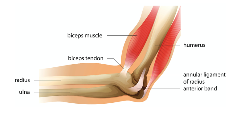
An elbow ligament tear refers to the damage or injury to one or more of the ligaments in the elbow joint. The elbow is supported by three primary ligaments: the ulnar collateral ligament (UCL), the radial collateral ligament (RCL), and the annular ligament.

Types of Elbow Ligament Tears
- Ulnar Collateral Ligament (UCL) Tear: The UCL is the most commonly injured elbow ligament. It is located on the inner side of the elbow and is often associated with sports like baseball and overhead throwing activities.
- Radial Collateral Ligament (RCL) Tear: The RCL is found on the outer side of the elbow. Tears in this ligament are less common.
- Annular Ligament Tear: The annular ligament surrounds the head of the radius bone and helps stabilize the joint. Injuries to this ligament can lead to problems with forearm rotation.
Factors Contributing to Elbow Ligament Tears :
The Elbow Ligament Tear occurs mostly in the following cases::
- Overuse: Repetitive stress on the elbow, as seen in sports like baseball, can lead to overuse injuries and ligament tears
- Trauma: Direct impact or sudden force on the elbow can result in ligament tears.
- Ageing: As people age, the ligaments can become weaker and more susceptible to injury.
- Occupational Factors: Certain professions that involve repetitive wrist and elbow movements can increase the risk of ligament injuries.

What are the common Symptoms of Elbow Ligament Tear ?
Common signs of Elbow Ligament Tear may include:
- Pain on the inner or outer side of the elbow, depending on the ligament affected.
- Swelling and tenderness.
- Reduced range of motion in the elbow.
- Weakness in the affected arm.
- Instability of the elbow joint.
- A popping or snapping sensation during movement.
How to diagnose Elbow Ligament Tear?
- A medical history and physical examination to assess pain, tenderness, and range of motion.
- Imaging studies such as X-rays to rule out fractures and MRI to visualise soft tissue damage.
- Sometimes, an arthrogram may be used, which involves injecting a contrast dye into the joint to enhance imaging.
- Stress tests, where the doctor may apply pressure to the joint to assess stability.
